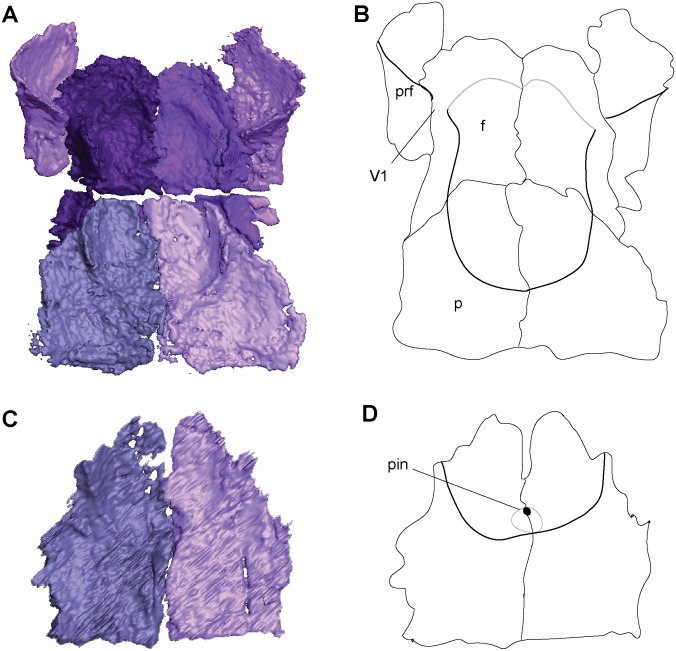
Fig 2. Ventral skull roof reconstructions and interpretive drawings of the descending flange of the prefrontals, frontals, and parietals.
Rhynchonkos stovalli (FM-UR 1039); (A, B) and Aletrimyti gaskillae, gen. et sp. nov. (FM-UR 1040); (C, D), in ventral view, based on select micro-CT images. Bold line indicates the descending flange of the prefrontal, frontal, and parietal. Grey line indicates a depression in the bone. Not shown to scale. Abbreviations: f, frontal; p, parietal; pin, pineal foramen; prf, prefrontal; V1, foramen for the ophthalmic branch of the trigeminal nerve.

