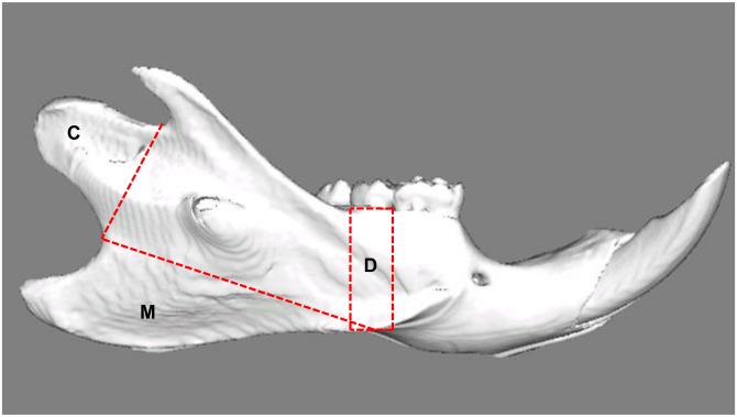
Fig 1. Lateral view of a reconstructed right hemimandible showing the volumes of interest.
C: Condyle; Selection is defined by the line connecting the two notches superior and inferior of this area. M: Attachment site of the superficial masseter muscle; the border line connects the notches ventrocaudal and craniodorsal to the mandibular angle. D: Alveolar cortical bone; the selection is defined by two lines drawn perpendicular to the lower border of the mandible, mesial and distal of the second molar.

