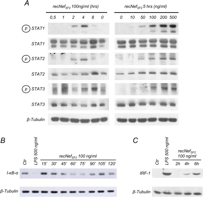
Fig 1. Nef treatment induces STATs tyrosine phosphorylation, I-B degradation and IRF-1 expression in BV-2 microglial cells.
(A) BV-2 cells were left untreated or incubated with 100 ng/ml wild type myristoylated Nef derived from HIV-1 SF2 strain (myr+NefSF2) for different times (left panel) or for 5 h with different amounts of myr+NefSF2 (right panel). Total cellular extracts were analyzed by Western Blot to evaluate STAT-1, -2 and -3 tyrosine phosphorylation or protein expression using specific antibodies as described in materials and methods. (B, C) Cells were incubated for the indicated time with myr+NefSF2 (100 ng/ml) or, as a positive control, with LPS (500 ng/ml) for 30’ in (B) and 6 h in (C). Whole cell lysates were analyzed by Western Blot for I-κB-α (B) or IRF-1 (C) expression. β-tubulin expression was used as an internal loading control. Results reported in the figure are from four independent experiments.

