
Figure 4. Possible binding sites of the different monoclonal antibodies in recent anti-Ebola virus cocktails.
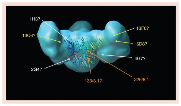
In light blue is a model of the fully glycosylated mucin-containing Ebola virus glycoprotein (GP), with a crystal structure of the mucin-deleted core drawn at center in ribbon representation. In the GP core ribbon model, GP1 subunits are blue and green while the GP2 subunits are colored gray. Antibody epitopes are roughly located by the site of amino acid mutations that confer escape. Two mAbs in the Marzi et al. cocktail, 226/8.1 and 133/3.16, are listed in orange [42]. mAbs in the MB-003 cocktail of Olinger et al. are listed in yellow [40]. mAbs in the Qiu et al. ZMAb cocktail are in white [41]. Although the identities of the amino acids in the mucin-like domain to which 13F6 and 6D8 bind are known, the location of these linear epitopes in the 3D space of the mucin-like domain is not yet known. Experimental mapping of antibody epitopes is a major goal of the Filovirus Immunotherapeutic Consortium.