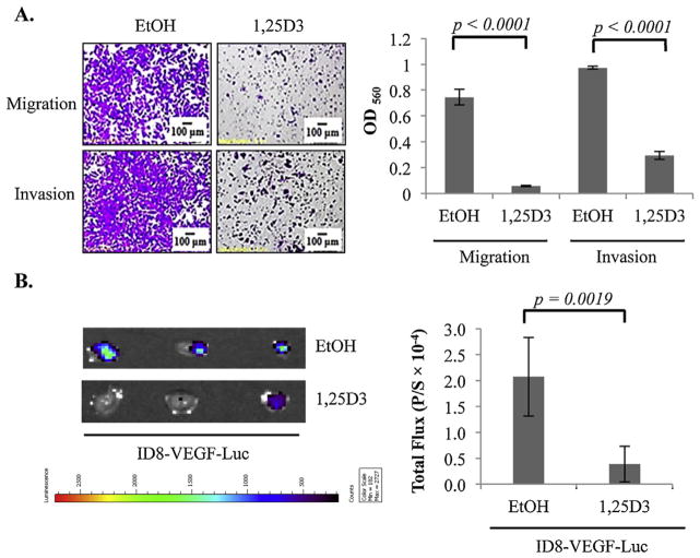
Fig. 3.
Suppression of EOC invasion to the omentum in mice by 1,25D3 through the epithelial VDR. (A) Suppression of migration and invasion of mouse epithelial ovarian tumor cells in transwell assays. ID8-VEGF-Luc cells were pre-treated with EtOH or 10−7 M 1,25D3. Migration and invasion were measured as in Fig. 1 for OVCAR3 cells. (B) Suppression of ID8-VEGF cell invasion into omenta in intact mice by 1,25D3 through the epithelial VDR. ID8-VEGF-Luc cells were pretreated with either EtOH or 1,25D3 (10−7 M) for 6 days. 5 × 10⁶ of treated cells were injected i.p. into mice (n = 5/group). After 18 h, omenta were isolated. Bioluminescence IVIS images were captured (left panels) and quantified (bar graphs, p/s = photons/second). The experiments were reproduced twice.

