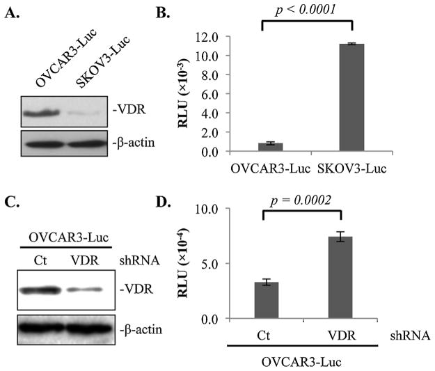
Fig. 4.
1,25D3-independent suppression of EOC invasion to the omentum by epithelial VDR revealed in ex vivo assays. (A) Immunoblotting analyses showing VDR protein expression levels in OVCAR3-Luc and SKOV3-Luc cells using β-actin as a loading control. (B) Normalized luciferase activities of OVCAR3-Luc and SKOV3-Luc cells invaded mouse omenta. (C) Immunoblotting analyses showing VDR protein expression levels in OVCAR3-Luc cell stably expressing control (Ct) or VDR (VDR) shRNA using β-actin as a loading control. (D) Normalized luciferase activities of OVCAR3-Luc cells expressing Ct or VDR shRNA invaded mouse omenta. For each data point, triplicate samples were analyzed. The experiments were reproduced twice.

