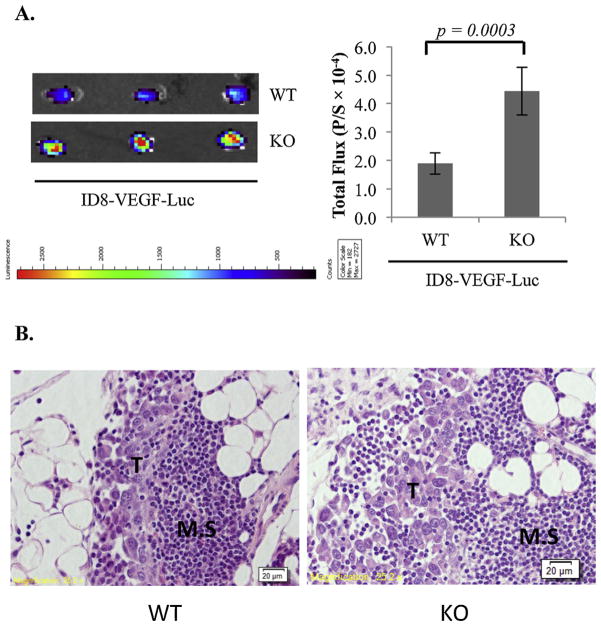
Fig. 6.
Suppression of EOC colonization to the omentum by stromal VDR in the ID8-VEGF syngeneic EOC mouse model. (A) 5 × 10⁶ ID8-VEGF-Luc cells were i.p. injected into WT (n = 5) and KO (n = 5) mice. 18 h later, omenta were isolated and bioluminescence IVIS images of the tumors were captured (left panels) and quantified (bar graphs, p/s = photons/second). (B) Sections of isolated omenta were subjected to hematoxylin and eosin stain. Tumors (T) established as metastatic are obvious in the milky spots (M.S.).

