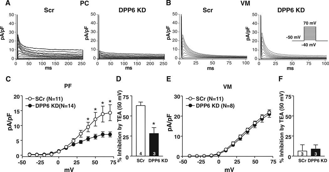
Figure 6. Effects of dipeptidyl peptidase-like protein-6 (DPP6) knockdown (KD) on Purkinje fiber cell (PC) and ventricular myocyte (VM) Ito.
Examples of Ito recordings from PC A, and VM B, infected with Adv-GFP-Scr (Scr) or Adv-GFP-DPP6 KD (DPP6 KD). Currents were obtained with 250- (PC) or 100-millisecond (VM) depolarizations at 0.1 Hz. C and E, Mean±SEM Ito density-voltage relations in Scr or DPP6 KD from PCs and VMs. D and F, Percentage inhibition by 10 mmol/L tetraethylammonium (TEA) of PC (D) or VM (F) Ito at 50 mV. *P<0.05, Scr vs DPP6 KD. GFP indicates green fluorescent protein.

