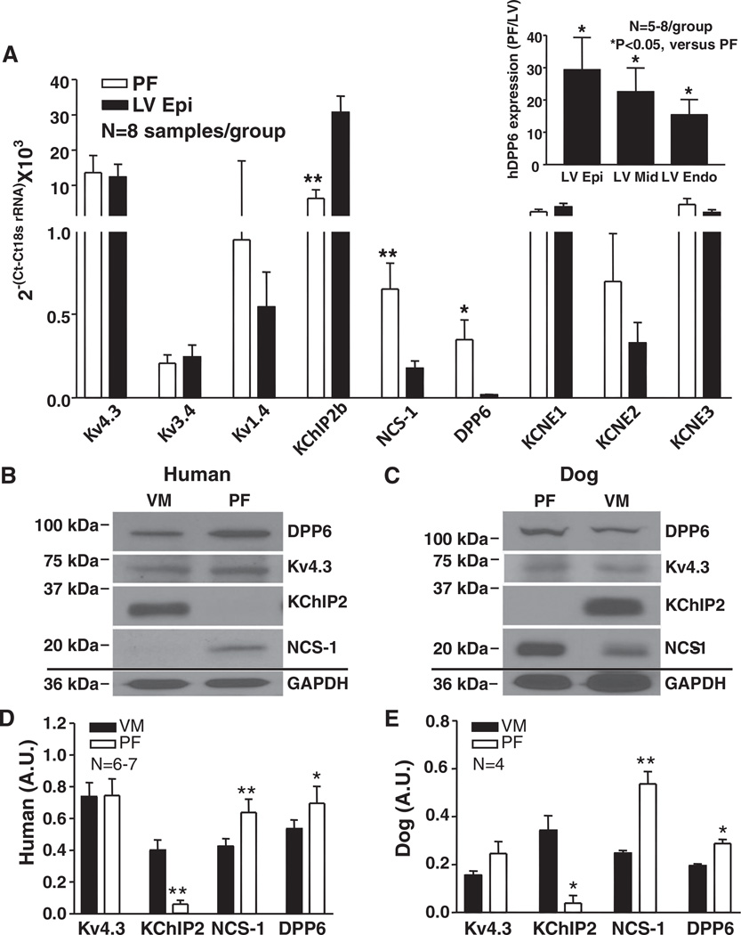
Figure 7. mRNA and protein expression of Ito subunits.
A, Ito subunit mRNA expression in human heart. Mean±SEM normalized results for Kv4.3, Kv3.4, Kv1.4, K+-channel interacting protein type-2 (KChIP2)b, neuronal calcium sensor-1 (NCS-1), dipeptidyl peptidaselike protein-6 (DPP6), KCNE1, KCNE2, and KCNE3. *P<0.05, **P<0.01, Purkinje fibers (PF) vs left ventricle (LV) epicardium (Epi). Inset, DPP6 mRNA expression in PF as ratio of LV Epi (n=8), midmyocardium (LV Mid) (n=5), and endocardium (Endo) (n=6). *P<0.05, PF vs LV layers. B and C, Representative Western blot results in ventricular muscle (VM) and PF membrane fractions from human (B) and dog (C) hearts. All blots shown were from 1 VM and 1 PF sample on the same membrane for each, which was stripped before each antibody was applied. D and E, Mean±SEM protein levels normalized to GAPDH. *P<0.05, **P<0.01, PF vs VM.

