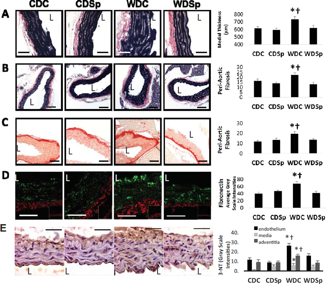
Figure 2.
Aortic remodeling in untreated western diet-fed mice (WDC) mice is prevented by MR antagonism (WDSp). (A) Representative micrographs show medial wall thickening, peri-aortic fibrosis by (B) VVG and (C) picrosirius red staining and (D) adventitial accumulation of fibronectin. (E) Immunostaining analysis for 3-nitrotyrosine staining. Abbreviations and symbols are the same as in Figure 2 legend. The vessel lumen is indicated by the letter L. Values are mean ± SE; n=5 per group.

