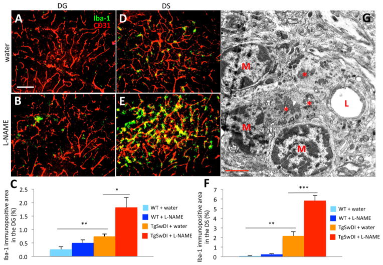
Figure 3. Hypertensive TgSwDI mice display increased vascular microgliosis.
Microgliosis was examined in tissues using an anti-Iba-1 antibody (green) and vessels were stained with an endothelial cell-specific anti-CD31 antibody (red). Compared to water-treated TgSwDI (A,D) and WT (not shown) groups, the DG (B) and DS (E) of hypertensive TgSwDI brains exhibited significantly more Iba-1 staining after 6 months of treatment (C,F; **p<0.01 normotensive TgSwDI vs. normotensive WT; *p<0.05 or ***p<0.001 normotensive vs. hypertensive TgSwDI; scale bar=100 μm; n=6–9/group). (G) The infiltration of microglia was observed by EM of brain capillaries in hypertensive TgSwDI samples. Capillaries laden with Aβ (*) were surrounded by infiltrating microglial nuclei (M, microglia; L, lumen; scale bar=2 μm).

