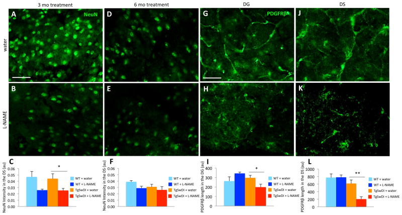
Figure 5. Hypertensive TgSwDI mice exhibit early neuron and pericyte loss.
Anti-NeuN antibody was used to identify neurons in the DS of normotensive (A,D) and hypertensive (B,E) TgSwDI mice. (C) Reduced NeuN intensity was observed in hypertensive TgSwDI mice after 3 months of L-NAME treatment (*p<0.05, n=3–9/group). (F) Since NeuN intensity was slightly decreased in normotensive TgSwDI animals after 6 months of treatment due to advanced age (9–10 months-of-age), there was no longer a significant difference between groups. Although NeuN intensity appeared reduced in the DS of hypertensive WT mice compared to normotensive WT mice after 3 and 6 months of treatment, neither change was significant (C,F). Similarly, anti-PDGFRβ antibody was used to examine pericyte coverage of vessels in normotensive (G,J) and hypertensive (H,K) TgSwDI mice. (I,L) PDGFRβ levels were significantly reduced in hypertensive TgSwDI mice in the DG and DS compared to control groups after 6 months of treatment (*p<0.05, **p<0.01; scale bars=50 μm; n=5–9/group).

