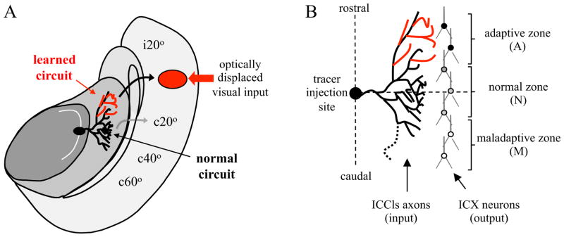
Figure 1. Experimental design.

A, Diagram of horizontal section through the R midbrain of a prism-adapted owl. Neurons in the central nucleus of the inferior colliculus (ICC) are tuned to distinct values of interaural time difference (ITD) and arranged topographically to form a map, indicated by the curved arrow located in the lateral shell of the ICC (ICCls). ITDs corresponding to ipsilateral space are represented in the rostral pole and ITDs corresponding to progressively more contralateral space towards the caudal pole. ICCls neurons project to the external nucleus of the inferior colliculus (ICX) where a complete map of auditory space is assembled. The axonal projection labeled by focal injection of anterograde tracer at c50 μs ITD is depicted. Black and red lines depict the normal and learned circuits. Major postsynaptic targets of these axons are CaMKII+ space-specific neurons in ICX (depicted in B). Curved arrows indicate the projection of CaMKII+ neurons to the OT where the auditory space map aligns with a visual space map. An ITD of 50 μs corresponds to 20° azimuth in visual space. In both ICX and OT, ipsilateral space is represented in the rostral pole and contralateral space progressively towards the caudal pole. Red arrow indicates the location of optically displaced visual input arising from a stimulus located 20° to the owl’s left (for illustration; actual prisms used were 19°). After full adaptation, depicted here, the learned circuit drives strong responses in ICX which are conveyed to the appropriate location in OT (black arrow), whereas the persistent normal circuit drives weak responses in ICX which are conveyed to the non-displaced location in OT (grey arrow). Following prism removal, the normal responses are re-expressed and the learned responses suppressed (see Figures 2 and 4). B, Microanatomical analysis in a prism-adapted owl. Contacts between tracer-labeled axons and CaMKII+ dendrites are distributed across the entire rostrocaudal extent of the axonal arbor, ~ 2mm, and with similar bulk density within the adaptive and normal zones. Nonetheless, in response to auditory stimulus and co-activation of these synapses, the postsynaptic cells located in the adaptive zone respond most strongly.
