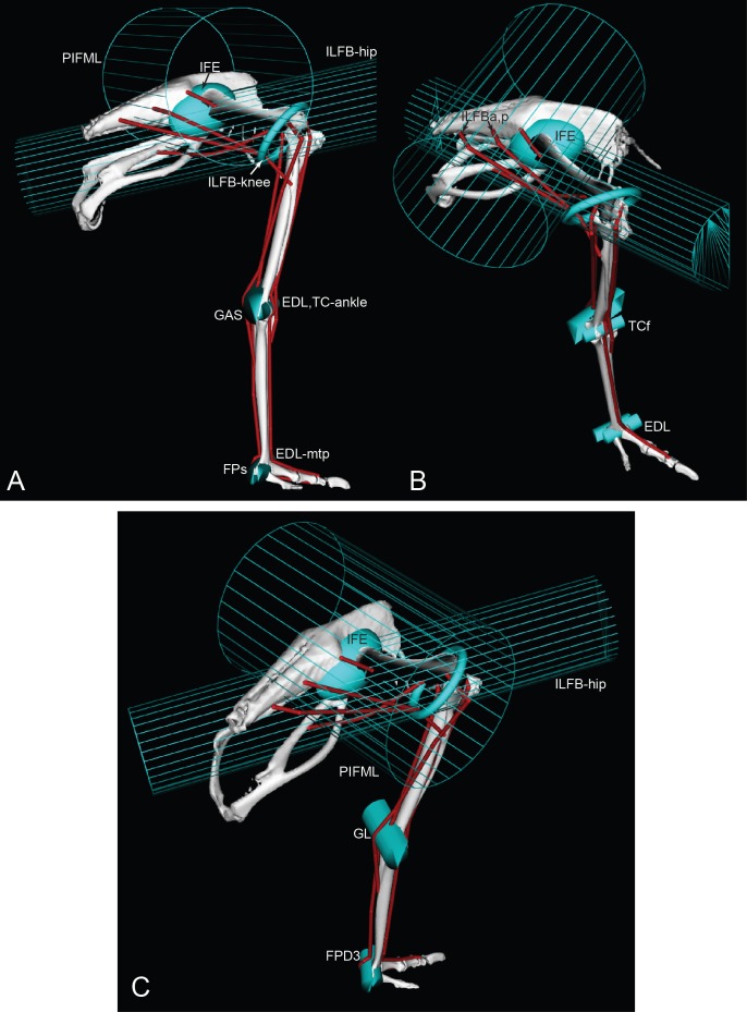
Figure 5. Ostrich musculoskeletal model: wrapping surface examples.
See Table 2 for muscle abbreviations. Lateral (A), craniolateral (B), and caudolateral (C) views of eight muscle wrapping objects (in blue), as half and whole cylinders, ellipses and a torus. The PIFML and ILFB wrapping surfaces are shown as meshes, for added clarity.

