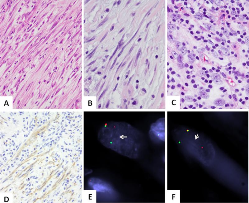
Figure 1. Pathologic findings of ROS1-rearranged IMTs.
Most cases showed a distinctiven spindle cell proliferation with long, slender cell processes, within a loose edematous stroma with scant inflammatory component (A. IMT6, 200x, B. IMT2, 400x). The only adult IMT case in this genomic group showed more plump cells with ill-defined cell borders, vesicular chromatin and distinctive nucleoli and more abundant lymphocytic inflammatory infiltrate (C. IMT3, 400x). ROS1 immunostaining highlights the long cell processed of lesional cells (D. IMT2, 200x). FISH showing balanced ROS1 (E) and TFG (F) break-apart signals (arrows; IMT6; red, centromeric; green, telomeric).

