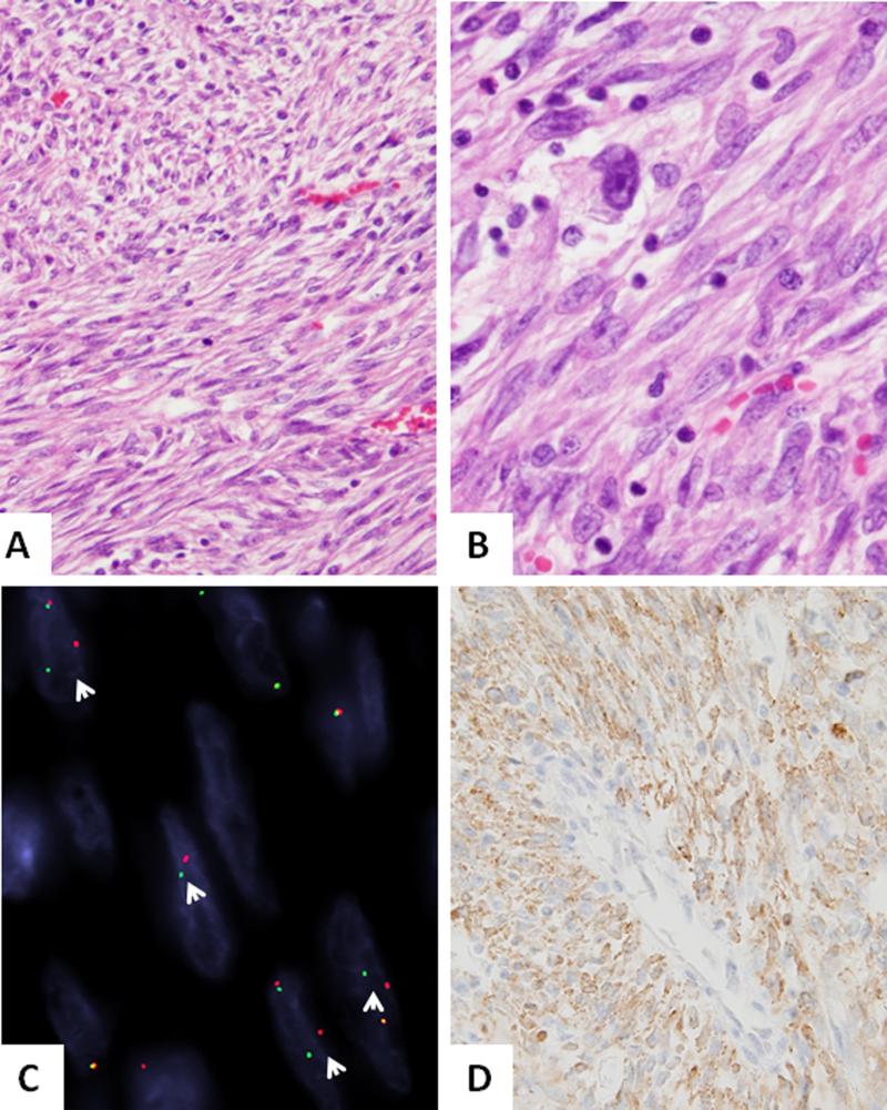
Figure 2. Novel RET-rearrangement in a pulmonary IMT (IMT7).
(A,B) A compact proliferation of relatively monotonous spindle cells arranged in intersecting long fascicles, with scant intervening stroma and mild inflammation (200x). Higher power depicts scattered pleomorphic cells with enlarged nuclei and prominent nucleoli (400x). C. FISH assay showing break-apart signals consistent with RET gene rearrangement (arrows, red centromeric, green, telomeric). D. ALK immunohistochemistry showing diffuse reactivity.

