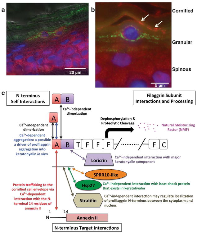
Figure 4. Profilaggrin N-terminus and stratifin co-localize in human epidermal granular cells.

Double label immunofluorescence was performed on fixed adult human skin using antibodies directed against the profilaggrin N-terminus (green) and stratifin (red). Panel (a) shows that stratifin is expressed throughout the epidermis, while profilaggrin is restricted to the granular layer and anuclear stratum corneum. (b) Shown is a vertical section of adult human skin immunolabeled with stratifin (red) and PNT (green) antibodies, with nuclei counterstained with DAPI. Stratifin is localized through the cytoplasm in spinous cells, but in the upper granular layer it is concentrated at the cell periphery where it co-localizes with PNT (orange labeling, arrows). Profilaggrin is also present in KHGs in the characteristic granular pattern, but shows little or no association with stratifin when present in the granular (profilaggrin) form. (c) Proposed biological functions of a calcium-dependent and calcium-independent protein interaction network for profilaggrin in human epidermis (based on the current study and the previous study by Yoneda et al., 2012).
