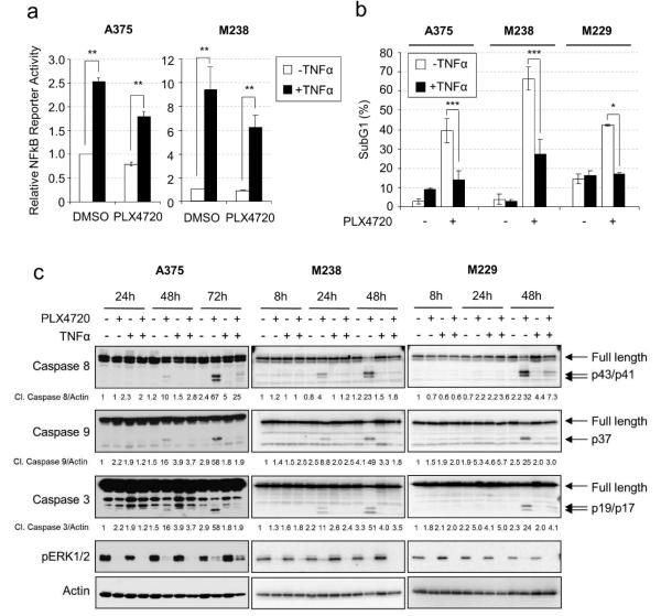
Figure 1. TNFα protects melanoma cells from RAF inhibitor-induced apoptosis.
a) Luciferase assays were performed as described in Material and Methods. Cells were treated with −/+ 50 ng/ml TNFα and −/+ 5 µM PLX4720 for 24 hours. Average relative luciferase activities from three experiments are shown. Error bars represent standard deviation. ** p<0.01. b) Cells were treated with −/+ 50 ng/ml TNFα and either −/+ 10 µM PLX4720 for 72 hours (A375) or −/+ 5 µM PLX4720 for 48 hours (M238, M229). Cells stained with PI for cell cycle analysis. Average percentages of sub-G1 populations from three experiments were quantified. * p<0.05; ** p<0.01; *** p<0.001. c) A375, M238 and M229 cells were treated with −/+ 50 ng/ml TNFα, −/+ 5 µM PLX4720 for 8-72 hours. Cells lysates were analyzed by Western blotting. Arrows indicate full length or cleaved products of caspases. Relative levels of cleaved caspases are indicated below the corresponding blots.

