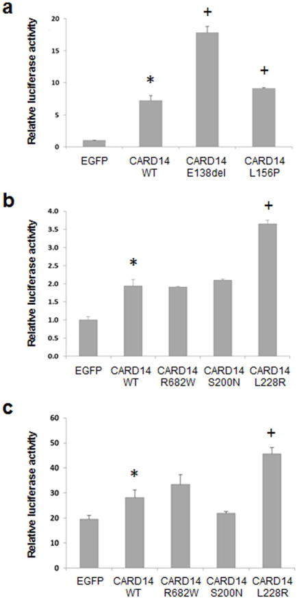
Figure 1. NF-κB activation by mutant CARD14 in cell culture systems in vitro.

The cells were transfected with wild type (WT) or mutant CARD14 cDNA constructs using Lipofectamine 2000 transfection reagent (Invitrogen, Carlsbad, CA), followed by assay of luciferase activity after 24 hours of incubation. The CARD14 cDNA (coding for 740 amino acids; GenBank BC018142) and EGFP cDNA as a control were cloned into pReceiver-M11 (Capital Biosciences, Rockville, MD) vector. (a) HEK293 cells were co-transfected with CARD14 constructs together with kB-Luc plasmid (kindly provided by Professor Yinor Ben-Neriah, Hebrew University, Jerusalem) and Renilla luciferase plasmid expression vector. Luciferase activity was measured using Dual-Luciferase® Reporter (DLT™) Assay System (Promega, Mullion, WI). (b) HeLa cells stably expressing NF-κB luciferase reporter (Signosis, Sunnyvale, CA) were co-transfected with the CARD14 constructs together with pRSV-galactosidase expression plasmid as a control of transfection efficiency. (c) The cultures as in (b) were supplemented after 24 hours of incubation with 20 ng/ml of recombinant human TNF-α (PeproTech, Rocky Hill, NJ). After an additional 24 hrs, luciferase activity was measured using Luciferase Assay System (Promega). The experiments were performed in triplicate cultures, and the values are expressed as mean + S.E. Statistical differences were evaluated by Student’s two-tailed t-test: *, p<0.05 as compared with EGFP as a control construct; +, p<0.05 as compared with CARD14 WT construct.
