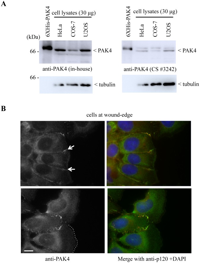
Fig 2. PAK4 does not localize to the leading edge of migrating cells.
A) Total lysates (30 μg per lane) extracted from HeLa, COS-7 and U2OS cells were probed for PAK4 by Western blotting using either an affinity purfied PAK4 antibody [14] or Cell Signaling (*3242). Recombinant purified His-tagged PAK4 (6XHis-PAK4) in lane 1 was loaded (10 ng) as a size control. B) Images showing the typical morphology of migrating U2OS cells at the wound-edge in a standard monolayer scratch assay. The cells were fixed with methanol and immuno-stained with anti-PAK4 and anti-p120-catenin. PAK4 enrichment at the cell-cell junction is indicated by white arrows (top panel), and the edge of tone of the lamellipodia is marked with a white dotted line (bottom panel). Scale bar: 5 μm.

