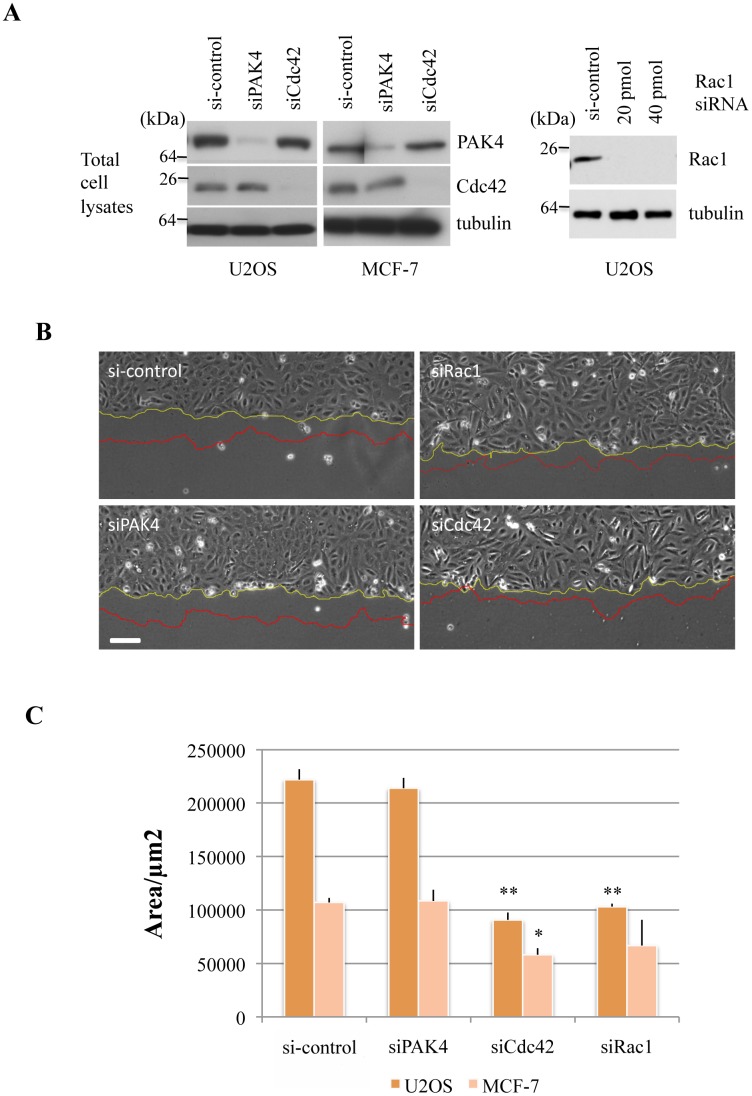
Fig 3. PAK4 loss does not affect collective migration rates of U2OS or MCF-7 cells.
A) (Left panel) U2OS or MCF-7 cells were transfected with siRNA directed to PAK4 or Cdc42 as indicated. The cell lysates (30 μg per lane) were probed for expression of PAK4, Cdc42 or tubulin. (Right panel) U2OS cells were transfected with siRNA directed to Rac1 as indicated and the lysates were probed for expression of Rac1 or tubulin. B) Low power images of the same area of the monolayer scratch wound are shown before and after 4h cell migration into the gap. The wound-edge is represented in yellow and red corresponding to the start and end of imaging respectively. C) Bar chart depicting the area covered over 4h after the scratch was applied by either the U2OS or MCF-7 cells, with standard error of mean. The area was calculated using ImageJ software. *P value < 0.05, **P value < 0.005. Scale bar: 50μm.

