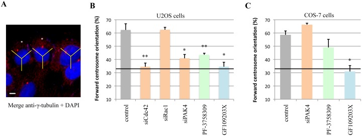
Fig 4. PAK4 regulates centrosomal reorientation in U2OS cells.
A) Diagram showing how cells were scored for forward centrosomal reorientation based on immuno-localization of γ-tubulin (prominent red dot) and the nucleus (blue). This was done using a 120° sector (indicated in yellow) centered on the nucleus, and cells with γ-tubulin placed within the forward sector facing the wound edge are scored (as marked with asterisks). The wound is toward the top of the figure and the image was acquired 1h post wounding. B) Graph showing the percentage of U2OS cells with forward centrosome orientation 1h post wounding. Cells were transfected with siRNA to Cdc42, Rac1 or PAK4 and left for 48h before analysis. Cells were also treated with either a PAK4 inhibitor PF-3758309 or a PKC inhibitor GF109203X for 1h prior to scratching. The data represents three independent experiments (N = 60) with standard error of mean. A random orientation of the centrosome gives a 33% baseline (solid black line). C) Graph showing the same analysis performed with COS-7 cells 1h post wounding. Cells were transfected 48h with siPAK4 or treated with inhibitors to PAK4 (PF-3758309) or PKC (GF109203X) for 1h before scratching. *P value < 0.01, **P value < 0.005. Scale bar: 10μm.

