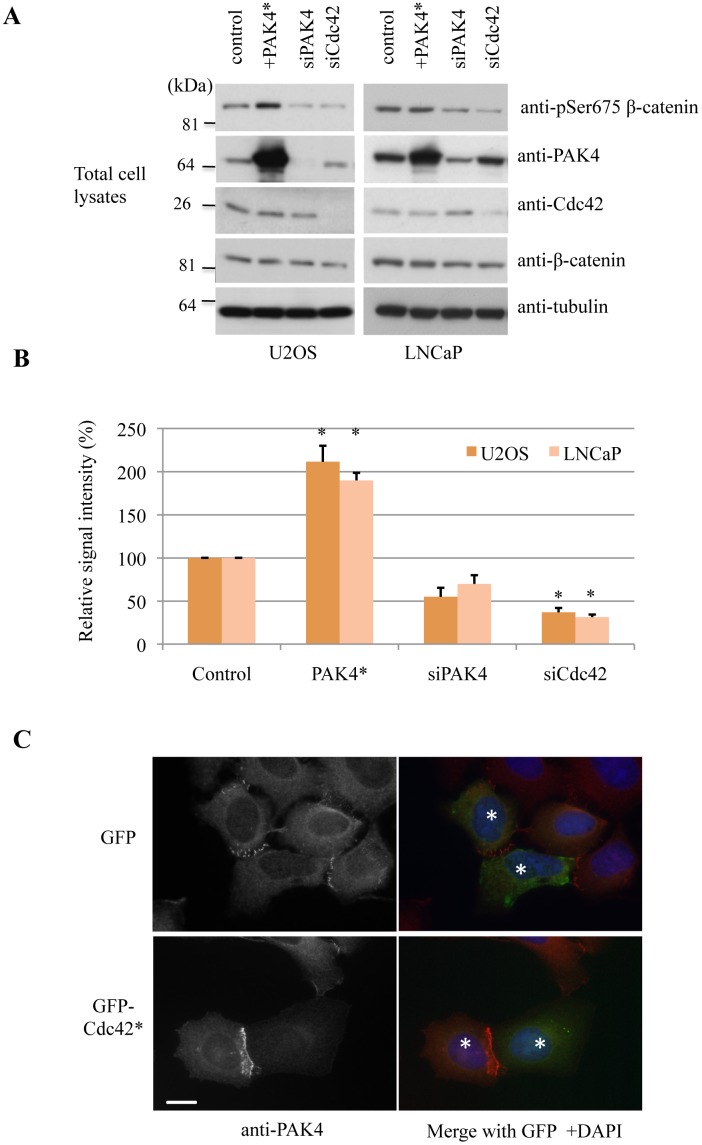
Fig 5. PAK4 phosphorylates β-catenin at Ser-675.
A) U2OS or LNCaP cells were transfected overnight with FLAG-PAK4 S445N (denoted PAK4*), or with PAK4 siRNA or Cdc42 siRNA for 48h. The cell lysates (30 μg per lane) processed for Western blotting are as indicated. B) The signal intensities corresponding to bands detected by anti-pSer-675 β-catenin or total anti-β-catenin were obtained from 3 independent experiments using ImageJ, averaged and plotted with standard error of mean. *P value < 0.05. C) U2OS cells were transfected with GFP or GFP-Cdc42(G12V) (GFP-Cdc42*), and fixed the following day. Cells were then immuno-stained for PAK4 and the nuclei were stained by DAPI. Two neighbouring cells in which the cell-cell junction was observed are denoted by asterisks. Scale bar: 10μm.

