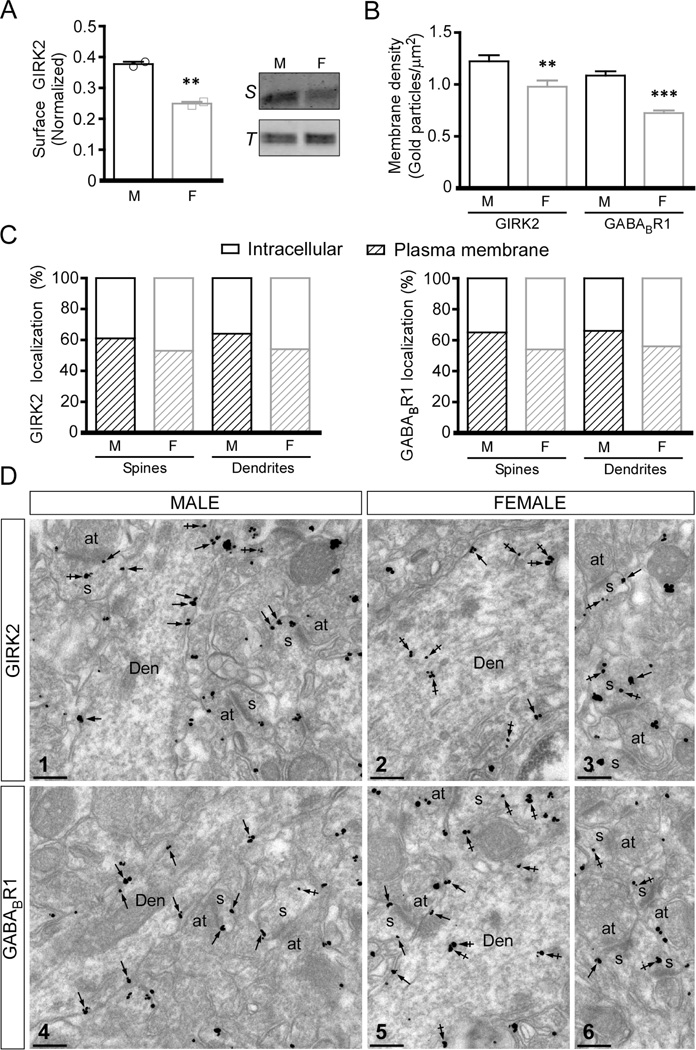
Figure 5. Subcellular localization of GIRK2 and GABABR1 in layer 5/6 pyramidal neurons of male and female mice.
A) Left: Quantification of surface GIRK2 protein levels in mPFC micropunches from adolescent male (M) and female (F) mice, with normalization to total GIRK2 protein (t2=13, P<0.01; n=2/group). Right: Representative immunoblots showing surface (S) and total GIRK2 (T) protein levels in mPFC micropunches from adolescent male and female wild-type mice. B) Plasma membrane-associated immunogold particle density for GIRK2 (Mann-Whitney rank sum test; T=5499, n=80; P<0.01) and GABABR1 (Mann-Whitney rank sum test; T=4380, n=80; P<0.001) in dendrites from adolescent male (M) and female (F) mice (n=4/sex) **,*** P < 0.01 & 0.001, respectively, vs. male. C) Distribution of GIRK2 and GABABR1 immunoparticles at the plasma membrane and intracellular sites in layer 5/6 PrLC pyramidal neuron spines and dendrites from adolescent male (M) and female (F) mice (n=4/sex), expressed as a percentage of total particles (n=4/sex). D) Representative images of GIRK2 (Image 1) and GABABR1 (Image 4) immunoreactivity in mPFC Layer 5/6 pyramidal neuron dendrites and spines from a male mouse. Immunoparticles for GIRK2 or GABABR1 were mainly detected along the extrasynaptic plasma membrane (arrows) of dendritic shafts (Den) and spines (s), and at low levels at intracellular sites (crossed arrows) in these compartments. (Images 2,3,5,6) Representative images of GIRK2 (Images 2,3) and GABABR1 (Images 5,6) immunoreactivity in mPFC Layer 5/6 pyramidal neuron dendrites and spines from a female mouse. Immunoparticles for GIRK2 or GABABR1 were detected along the extrasynaptic plasma membrane (arrows) of dendritic shafts (Den) and spines (s), and more frequently observed at intracellular sites (crossed arrows) in these compartments. Abbreviation: at, axon terminal. Scale bars: 0.2 µm.

