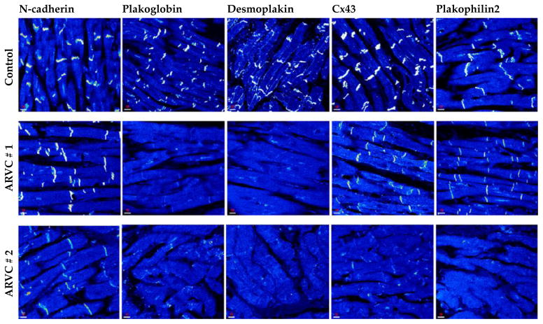
Figure 2.
Representative confocal immunofluorescence images of control myocardium and myocardium from two patients with autosomal dominant ARVC. Specific immunoreactive signal for plakoglobin was depressed at cell-cell junctions in the great majority of cases regardless of the underlying pathogenic mutation. Signal for desmoplakin and plakophilin2 varied, while signal for N-cadherin was always present and indistinguishable from controls. The majority of cases examined showed gap junction remodeling as evidenced by decreased junctional signal for Cx43 (reproduced from Asimaki et al. NEJM 2009; 360:1078).41

