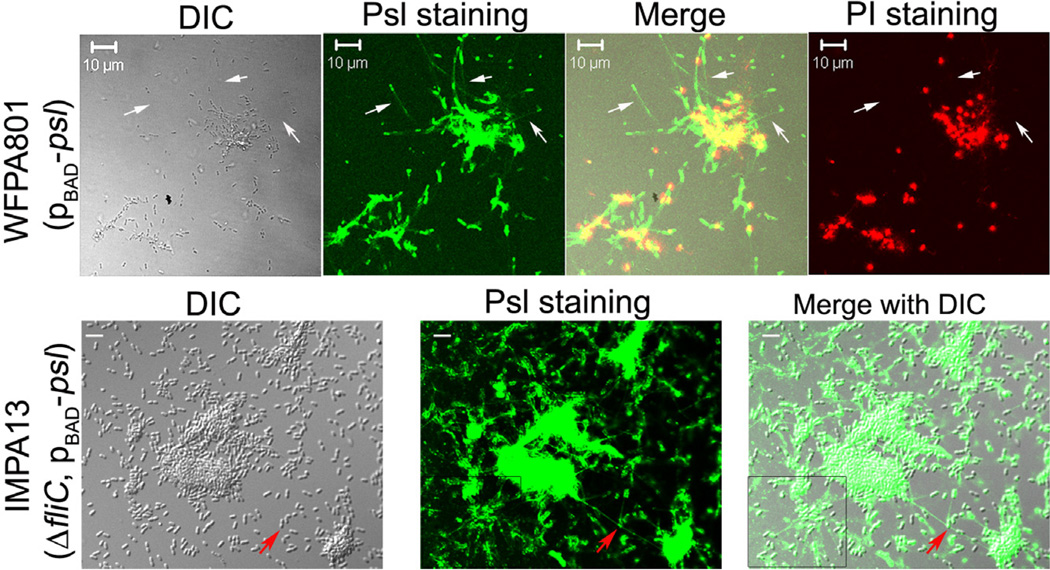
Fig. 7.
Psl tracks were detected after bacterial cells attached on a glass slip. Shown are surface-attached bacterial cells of WFPA801 and IMPA13 stained by HHA-FITC and/or propidium iodide (PI). White arrows pointed out the HHA-FITC stained Psl tracks (green) following bacterial cells. Red fluorescent signals were PI stained DNA of dead/dying bacteria. The red arrow indicates a Psl-fibre strand connecting two microcolonies. The boxed area showed the web-like radial pattern Psl tracks. Grey panel was the corresponding DIC image. Scale bar: 10 mm for WFPA801, 5 µm for IMPA13.

