
Fig. 2.
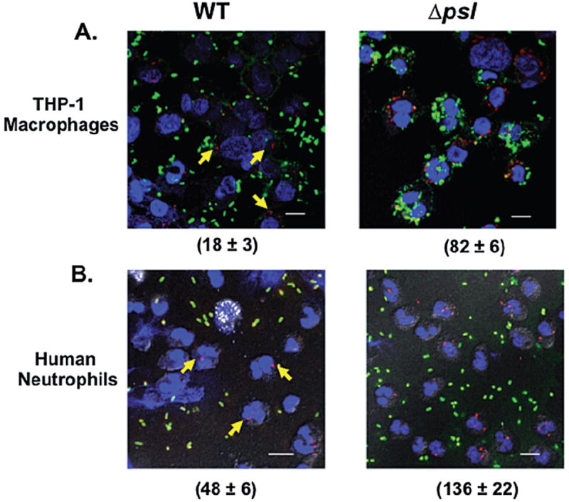
The presence of Psl reduces the uptake of P. aeruginosa by human phagocytic cells. Immunofluorescence images of (A) THP-1 cells and (B) human neutrophils, infected with pre-opsonized WT or a psl-deficient strain of P. aeruginosa. Bacteria were incubated with neutrophils at an moi of 5:1 and processed as described in Experimental procedures. Attached bacteria exposed to the antibodies before permeabilization stained green. Those exposed to the antibodies only after permeabilization stained red (internalized bacteria). Blue (DAPI) fluorescence was used to stain the nuclei of THP-1 cells and neutrophils (60× oil objective). Arrows indicate the internalized bacteria in the PAO1 samples. Scale bar is equal to 10 μm. The number (mean ± SD) of internalized bacteria is indicated in parentheses and is representative of 100 infected cells (neutrophils and macrophages) examined from triplicate coverslips.