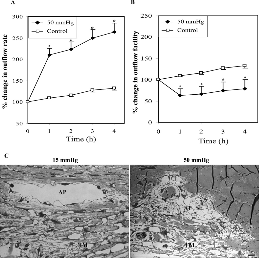
Fig. 1.
Elevated IOP in enucleated porcine eyes increases aqueous outflow rate and decreases aqueous outflow facility. A and B shows percent change in aqueous outflow rate and aqueous outflow facility from baseline in eyes perfused at 50 and 15 mm Hg, respectively. The eyes perfused at 50 mm Hg showed a significant increase in aqueous outflow rate (µl/min) from the base line value compared to eyes perfused at 15 mm Hg (A). On the other hand, the aqueous outflow facility (µl/min/mm Hg) was found to be decreased significantly from base line facility in eyes perfused at 50 mm Hg compared with eyes perfused at 15 mm Hg (B). Values represent mean ± standard error. n = 7, *p < 0.05, **p < 0.01. C. Porcine eyes perfused at 50 mm Hg for 5 hours revealed collapsed aqueous plexi (AP) and disorganized/compressed TM compared with eyes perfused at 15 mm Hg, based on histological evaluation by transmission electron microscopy. Bar indicates 10µm image magnification.

