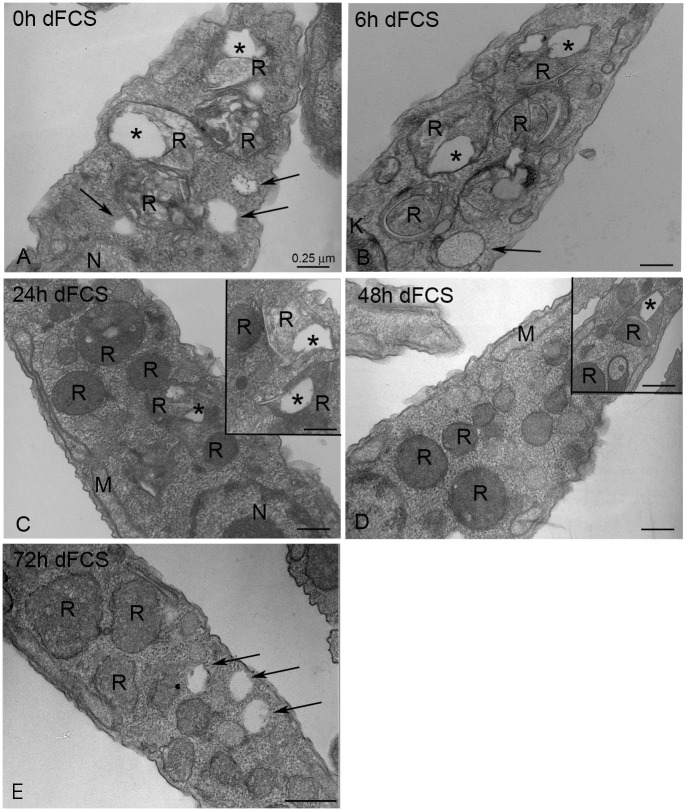
Fig 1. Transmission electron microscopy of epimastigotes cultivated in LIT medium supplemented with 10% dFCS.
(A) 0 h—Reservosomes show many lipid inclusions in their lumen (asterisk). (B) After 6 h of lipid starvation, reservosomes filled with lipid inclusions could still be observed, but after 24 h (C), 48 h (D) and 72 h (E), an expressive decrease in reservosome lipids was registered. The insets show that there were reservosomes with typical lipid inclusions even after 24 to 72 h of starvation. The arrows point to lipid bodies throughout the cytoplasm. K- Kinetoplast, M—Mitochondrion, N—Nucleus, R- reservosomes. Bars correspond to 0.25 μm.

