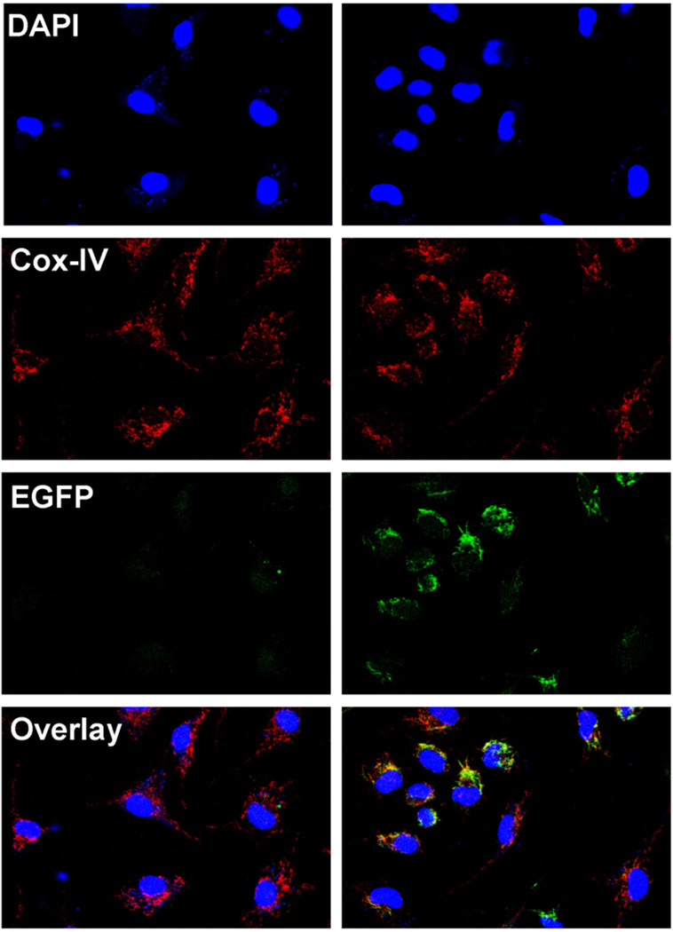
Fig. 4. Transgenic Grx2a-EGFP fusion protein localizes to mitochondria.
Confocal microscopy images of peritoneal macrophages isolated from wildtype (Wt) and transgenic CD68-Grx2aTg (Tg) mice and labeled with the mitochondrial marker Cox-IV (red) and the nuclear dye DAPI (blue). EGFP expression is shown in green. Image overlay shows co-localization of EGFP with the mitochondrial marker Cox-IV.

