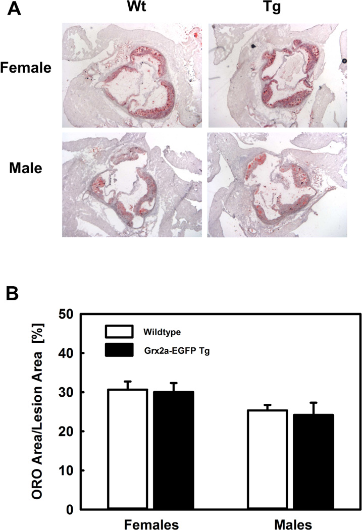
Fig. 7. Increased expression of Grx2a in monocytes and macrophages did not affect HFD-induced atherosclerotic lesion formation.
(A) Representative image of Oil red O (ORO)-stained sections of the aortic root from female and male LDLR−/− (wildtype; Wt) and transgenic Grx2aMacLDLR−/− mice (Tg) fed a HFD for 14 weeks. (B) Quantification of atherosclerosis in the aortic root wildtype and transgenic Grx2aMacLDLR−/− mice. There was no statistically significant difference in lesion size between LDLR−/− mice (open bars) and Grx2aMacLDLR−/− transgenic mice (closed bars). Females: n=9 per group; Males: n=12 per group.

