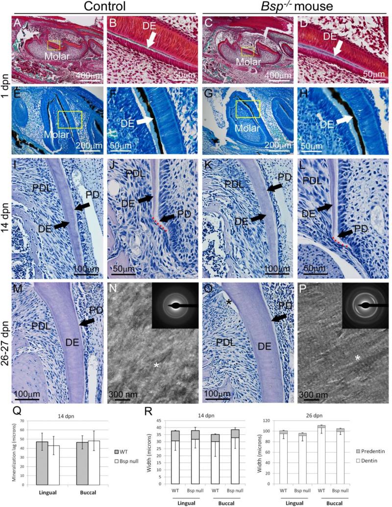
Figure 6. Loss of BSP does not affect molar dentin formation or mineralization.
Compared to controls, initiation of dentin (DE) mineralization is not delayed in Bsp−/− mouse molars by (A-D) Goldner's trichrome staining or (E-H) von Kossa staining, of undecalcified sections at 1 dpn. Yellow boxes in A, C, E, and G indicate areas shown under higher magnification in B, D, F, and H, respectively. The lag in mineralization between predentin (PD) matrix secretion (pale, whitish) and mineralization to dentin proper (light pink-purple) at the apical root tip is not delayed in Bsp−/− mice compared to control (J vs. L, and Q). No differences in the widths of root dentin and predentin at (I, K) 14 dpn or (M, O) 26 dpn in Bsp−/− vs. WT are observed, as confirmed by statistical analysis in R (n=4-6 samples per genotype for all ages). (N, P) TEM micrographs of unstained, natively mineralized dentin sections from 27 dpn mice show normal characteristics in both WT and Bsp−/− litter mates. In both micrographs, regions of collagen banding are evident (asterisk), indicating preferential gap zone mineral deposition. Electron diffraction (insets) in both WT and Bsp−/− molars show oriented arcs indicating preferential alignment of the hydroxyapatite (002) plane with the collagen fibril axis.

