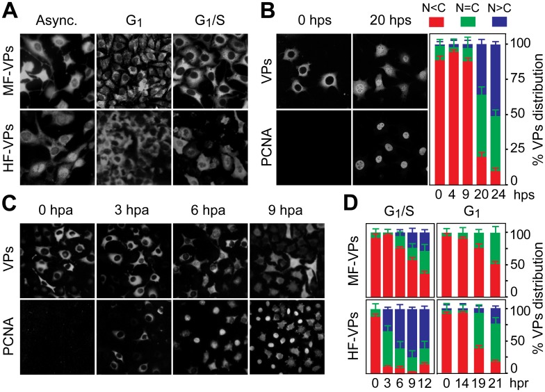
Fig 1. Cell cycle regulation of the nuclear translocation of MVM capsid proteins.
A. MVM capsid proteins (VPs) are excluded from the nucleus at G0/G1. Microscopy analysis of mouse (MF-VPs) and human (HF-VPs) fibroblasts stably expressing VPs fixed as asynchronous cultures (async.), synchronized by density arrest (G1), or by isoleucine deprivation/aphidicolin (G1/S). B. Kinetic of VPs nuclear transport in quiescent (G0) mouse fibroblast induced into cycle by serum. Left, cells stained with the α-VPs and PCNA antibodies. Right, average percentages with standard errors from three experiments of VPs subcellular distribution at the indicated hps. C. VPs nuclear transport is allowed at S phase. PCNA and VPs subcellular distribution in synchronous MF-VPs at the indicated hpa. D. VPs phenotypes scored in mouse and human fibroblasts hours post-release (hpr) of synchronization by aphidicolin (G1/S), or by density arrest (G1). Bars represent average values with standard errors from three or four experiments. (N<C (red): predominantly cytosolic, N = C (green): equal distribution between cytoplasm and nucleus, N>C (blue): predominantly nuclear).

