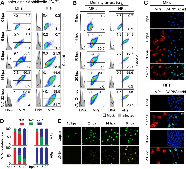
Fig 3. Cell cycle dependence of VPs expression and capsid assembly in the synchronous MVM infection of mammalian fibroblasts.
VPs expression and capsid formation in infected mouse (MFs) and human (HFs) fibroblasts analyzed by flow cytometry (A, B), or IF (C, D). Cells were synchronized by (A) isoleucine deprivation+aphidicolin (G1/S), or by (B) culturing to high density (G1), and stained for DNA content with DAPI (histograms: blank for mock, grey-filled for infected), VPs and Capsid (biparametric dot plots), at the indicated hours post-arrest releases (hpa or hpc, respectively). The percentage of cells in the corresponding gates is shown. CC, cell count. (C) Nuclear transport and assembly of MVM capsid subunits in synchronously infected mammalian fibroblasts. The figure shows representative fields of VPs and Capsid localized by double IF in MVM infected mouse and human fibroblats at the indicated hours post-release of the isoleucine/aphidicolin (G1/S) or growth to confluence (G1) cell cycle arrests. Scale bar 25 μm. (D) VPs subcellular distribution in synchronously infected mouse and human fibroblasts at the indicated hours post-release of the G1/S (hpa) or G1 (hpc) arrests. Average values with errors from four experiments are shown. (E) Synchronously infected MFs at G1/S stained for assembled capsids and viral DNA synthesis by FISH-hybridization (vDNA) along hpa. The figure shows representative confocal fields of cells from three experiments. Scale bar 10 μm.

