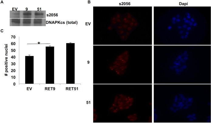
Fig 2. Phosphorylation of DNA-PKcs at s2056 is elevated in RET 9 and RET 51 cells.
A) Western blot analysis shows phospho-s2056 (ps2056) (460 Kda) to be elevated in RET9 and RET 51 cell lysates. Total DNA-PKcs was used as loading control. B) Immunocytochemistry of cells plated in chamber slides and stained for ps2056 (red) and Dapi as counterstain (blue). C) ICC revealed a significant increase in ps2056 located in the nuclei of RET 9 and RET 51 cells compared to EV. * p ≤ 0.05, error bars = s.d.

