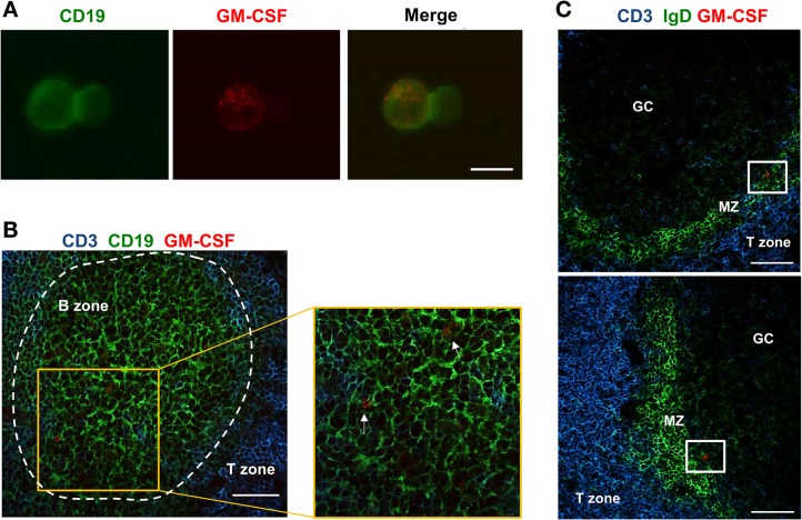
Fig 3. Human IRA B cells reside within the follicles.
A) Cell suspensions from tonsils were seeded onto poly-L-lysine coverslips and fixed with formaldehyde. Cells were incubated for 1 h with anti-CD19 (Green) and anti-GM-CSF (Red). The merge panel shows the co-localization of GM-CSF and CD19. The picture is representative of three different subjects. Scale bar = 10 μm. B) 8-μm tonsil tissue sections were fixed with formaldehyde and stained using anti-CD3 (T cell area, Blue), anti-CD19 (B cell area, Green) and anti-GM-CSF (Red). The enlargement shows the presence of GM-CSF+ cells within B cell follicles (white arrows). The panel is representative of 3 independent experiments using different tonsils where many sequential sections were screened (n>5). Scale bar = 50 μm. C) 8-μm tonsil tissue sections were fixed with formaldehyde and stained using anti-CD3 (T cell area, Blue), anti-IgD (mantle zone, Green) and anti-GM-CSF (Red). The panel is representative of 3 independent experiments using different tonsils where many sequential sections were screened (n>5). Scale bar = 50 μm.

