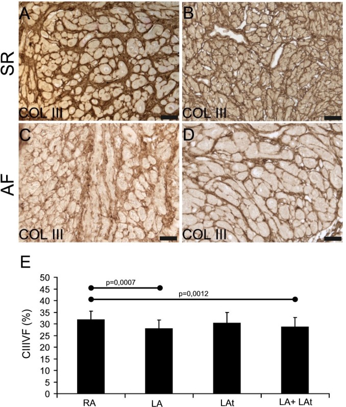
Fig 2. Collagen III in the atrial myocardium.

A-D: Immunohistochemical reaction shows collagen III in endomysial and partly also perimysial extracellular matrix. In atria of both patient groups (SR in A, B and AF in C, D) it is possible to detect higher (A, C) as well as lower (B, D) amount of collagen III-positive ECM. Immunoperoxidase reaction with DAB as a substrate (brown precipitate). No nuclear counterstaining. For all images scale bar = 50μm. (E) A graph showing the result of quantification of collagen III volume fraction (CIIIVF) in atrial myocardial samples. A comparison between different anatomical locations is shown: right appendage–RA (n = 37), left appendage–LA (n = 19), left atrium—LAt (n = 9), LA+LAt (n = 28).
