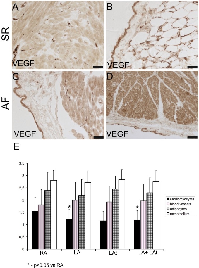
Fig 4. Expression of VEGF in atrial myocardium from patients with sinus rhythm (SR) and atrial fibrillation (AF).

A, B: Atrial samples from patients with SR. (A) A strong immunoreactivity for VEGF is localized to the capillaries, while cardiomyocytes display rather low level of VEGF expression. (B) High level of VEGF immunoreactivity in mesothelial cells and in adipocytes of epicardium. C, D: Atrial samples from patients with AF. (C) A strong immunoreactivity for VEGF is localized to mesothelium. There are VEGF-positive capillaries and moderately positive cardiomyocytes in the atrial myocardium. (D) A strong VEGF immunoreactivity in the myocardium (mainly cardiomyocytes) and in mesothelial cells. Scale bar in A-D = 50μm. (E) A graph showing a result of semiquantitative analysis of VEGF immunoreactivity in the atrial samples from all patients as described in Methods (score 0–3). A comparison between different structures from different anatomical locations is shown: RA–right appendage, LA–left appendage, LAt–left atrium.
