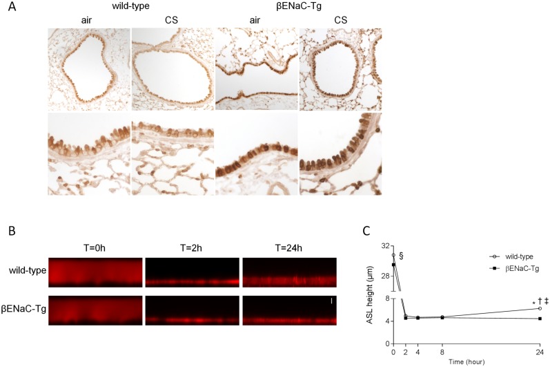
Fig 1. Overexpression of βENaC and reduced airway surface liquid height in βENaC-Tg mice.
Immunolocalization of βENaC in airways from WT and βENaC-Tg mice. (A) Representative images of βENaC immunostaining of lung sections from WT and βENaC-Tg mice that were exposed to air or CS for 8 weeks. n = 5 per group. Dysregulation of steady state airway surface liquid (ASL) height on airway epithelia from βENaC-Tg mice under thin film conditions. Representative confocal images (B) and summary of measurements of airway surface liquid height (C) at t = 0, 2, 4, 8 and 24h after mucosal addition of 20 μl of PBS containing Rhodamine dextran to primary tracheal epithelial cultures from βENaC-Tg mice and WT littermates. Scale bar, 7 μm. n = 4 experiments per group. *p<0.001 compared to βENaC-Tg; §p<0.001 for t = 0h compared to all other time points within the same genotype; †p<0.05 for t = 24h wild-type compared to t = 2h wild-type; ‡p<0.005 for t = 24h wild-type compared to t = 4h and 8h wild-type.

