
Figure 1. CK2α and CK2β expression by immunohistochemistry in FL, BL and DLBCL.
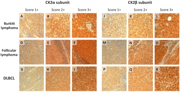
Representative immunohistochemical features of the considered lymphoproliferative lesions. Both BL (A-C; J-L), FL (D-F; M-O), and DLBCL (G-I; P-R) displayed consistent positivity for the regulatory and catalytic CK2 subunits. In all the considered lymphoma subtypes, variable immunohistochemical positivity (from score 1+ to score +3) was observed (H&E and immunoperoxidase stain; original magnification, x20).