
Figure 1. The elevated expression of S100A6 was detected in mRNA, protein and tissue of ccRCC samples.
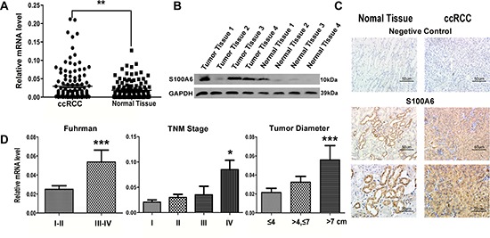
The elevated S100A6 correlated with ccRCC pathologic and clinical characteristics. (A) Real-time PCR analysis of S100A6 mRNA levels in ccRCC (n = 129) and adjacent non-tumor tissues (n = 129). mRNA levels were normalized to PPIA expression. (B) Protein expression of S100A6 in paired samples of ccRCC and adjacent non-tumor tissues by Western blot analysis. GAPDH served as loading control. (C) Representative figures of the immunohistochemistry specimens of S100A6 in ccRCC and matched non-tumor tissues, and with negative controls in each group. (D) The total 129 ccRCC cases were divided into sub-groups according the Fuhrman Grade, TNM Stage and the maximum diameter of tumor. The Real-time PCR assay showed the relative S100A6 mRNA levels in each groups. All the Relative mRNA data were expressed as mean ± SD and comparisons were performed using Student's t-test. *p < 0.05; **p < 0.01; ***p < 0.001.