
Figure 4. CAPE suppressed tumor growth of LNCaP 104-R1 xenografts.
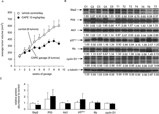
(A) LNCaP 104-R1 cells were injected subcutaneously into athymic mice to form tumors. After 14 weeks, the average tumor volume exceeded 150 mm3. The mice were then separated into control group and CAPE treatment group. Control group contained 6 mice and 8 tumors, while CAPE treatment group contained 6 mice and 9 tumors. CAPE (10 mg/kg/day in sesame oil) or vehicle (sesame oil) was administered by gavage starting from 14th week after cancer cell injection and was shown as 1st week for gavage in figure. Tumor volume and body weight of mice carrying 104-R1 xenografts were measured weekly. Tumor volume was shown as volume plus standard error (SE). Mice body weight in two groups did not show significant difference. (B) Protein expression of Skp2, p53, Akt1, p27Kip1, cyclin D1, and Rb in LNCaP 104-R1 tumors from control group or CAPE treatment group was assayed with Western blotting assay. α-tubulin was used as loading control. (C) The average expression level of Skp2, p53, Akt1, p27Kip1, cyclin D1, and Rb proteins in CAPE-treated LNCaP 104-R1 tumors was compared to those in tumors from control group. Asterisk* represents statistically significant difference p < 0.05 between the two groups.