
Figure 1. Release of PGE2 from γ-irradiated cells.
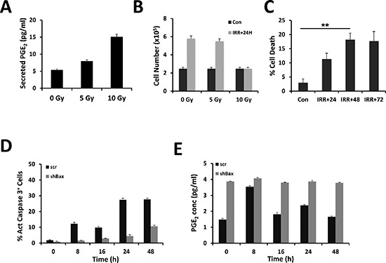
U251 cells were irradiated in serum-free medium at the indicated dose. PGE2 secretion was measured in supernatants after 24 h. The concentration of PGE2 (pg/ml) released from irradiated U251 cells was determined using an ELISA (see materials and methods) (A). Cell viability was determined by trypan blue exclusion using the Countess automatic cell counter (Life Technologies), 24 h after irradiation of U251 cells (B). Cell death was estimated as above at 24 h, 48 h and 72 h post-irradiation (C). U251 cells were transduced with shRNA encoding viral particles [either encoding for a non-relevant shRNA (scr) or shRNA directed against Bax mRNA]. Cells were irradiated in serum-free medium at 10 Gy; harvested at the indicated time points, fixed and labeled with active caspase 3 antibody coupled to a fluorescent secondary antibody. The percentage of labeled cells was assessed by flow cytometry (D). The corresponding PGE2 secretion was measured in supernatants during 48 h after irradiation at 5 Gy (E). Please note that in the latter experiments, the secretion of PGE2 was decreased in scr-treated U251 compared to untreated cells (compare A and E).