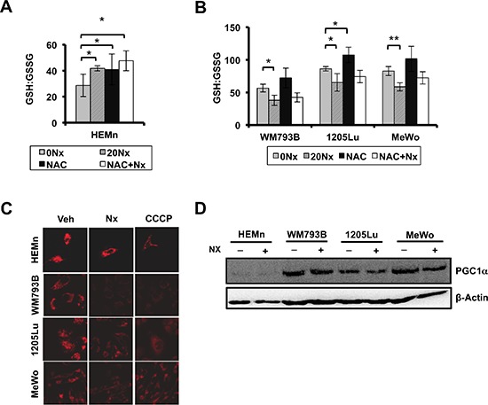
Figure 3. NexrutineR induces oxidative stress selectively in melanoma cells.

(A) Effect of NexrutineR treatment (vehicle, 20 μg/ml; 18 h) and NAC pre-treatment (5 mM; 1 h) on ratio of reduced (GSH) to oxidized (GSSG) intracellular glutathione in HEMn melanocytes, determined using luminescence-based assay. (B) Effect of NexrutineR treatment (vehicle, 20 μg/ml; 18 h) and NAC pre-treatment (5 mM; 1 h) on ratio of reduced (GSH) to oxidized (GSSG) intracellular glutathione in melanoma cells, determined using luminescence-based assay. (C) Evaluation of mitochondrial membrane potentials by confocal microscopy and incubation with TMRM potentiometric dye after NexrutineR (20 μg/ml; 3 h), 40X magnification. CCCP was used as a positive control for membrane uncoupling. (D) Protein level of PGC1α after NexrutineR (20 μg/ml; 18 h), evaluated by western blotting. Significance values; *indicates p ≤ 0.05; **indicates p ≤ 0.01.
