
Figure 2. Downregulation of Wnt5a in SGC-7901 cells is necessary for EGF-induced EMT.
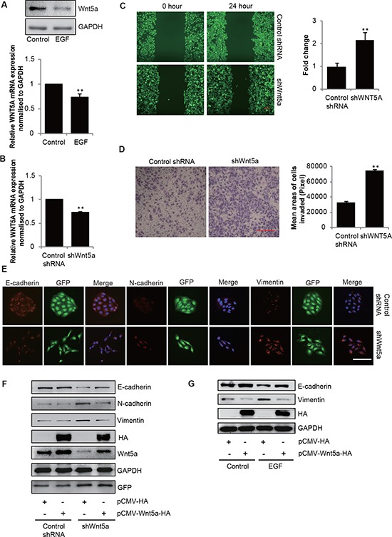
(A) Immunoblotting and qPCR analyses of Wnt5a mRNA and protein expressions in SGC-7901 cells that were incubated in the absence or presence of EGF (20 ng/mL) for 48 h. Data are presented as mean ± SD of 3 determinations, **P < 0.01 in the cultures with EGF relative to the cultures without EGF. (B) qPCR analyses of Wnt5a mRNA expression in SGC-7901 cells stable transfected with shRNA for Wnt5a. **P < 0.01 in the shWnt5a cells relative to the control group. (C) Wound healing assay and (D) transwell migration assay of control and Wnt5a knockdown cells. Scale bar, 100 μm. Data are presented as mean ± SD of 3 determinations, **P < 0.01 in the Wnt5a knockdown cells relative to control cells. (E) Representative immunofluorescence images of control and Wnt5a knockdown cells stained for E-cadherin, Vimentin and N-cadherin. Scale bar, 100 μm. (F) Immunoblotting analyses of E-cadherin, N-cadherin and Vimentin in control and Wnt5a knockdown cells transfected with vector (control) or HA-Wnt5a. (G) Cells were transfected with empty vector or HA-tagged Wnt5a, and then incubated with 20 ng/mL EGF for 48 h, the total cellular proteins were extracted and analyzed for expressions of E-cadherin by immunoblotting assays.