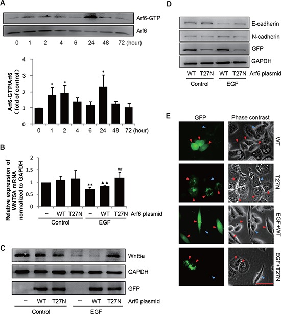
Figure 3. Arf6 mediates the EGF-induced EMT by down-regulating Wnt5a.

(A) SGC-7901 cells were treated with 20 ng/mL EGF for indicated times, and analyzed for Arf6 activity by pulldown and immunoblotting assays. Data are presented as mean ± SD of 3 determinations, *P < 0.05 cultures with EGF relative to the cultures without EGF. (B–E) SGC-7901 cells were transfected with Arf6-WT or Arf6-T27N plasmids, then treated with 20 ng/mL EGF for 48 h, (B) mRNA and (C) protein level of Wnt5a, (D) E-cadherin and N-cadherin were detected by qPCR and immunoblotting assay separately. Data are presented as mean ± SD of 3 determinations, **P < 0.01 in cultures with EGF relative to the cultures without EGF. ▲▲P < 0.01 in the empty vector-expression cells cultured with EGF relative to the cultures without EGF. ##P < 0.01 in the cells transfected with the Arf6 T27N expression vector treated with EGF relative to the cells transfected with empty vector treated with EGF. (E) The changing shape of cells by EGF was captured by phase-contrast microscopy. Scale bar, 50 μm. Arrows in the panels point to morphology appearance of cells transfected with or without GFP.
