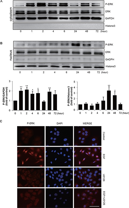
Figure 4. EGF induces P-ERK transportion to nucleaus.

(A & B) SGC-7901 cells were incubated with EGF 20 ng/mL for indicated times, then the extracts of plasma and nuclear section of SGC-7901 cells were subjected to immunoblotting analysis to detect the location and expression of P-ERK separately. GAPDH and Histone 3 are used for detecting cytoplasm and nucleus part. *P < 0.05, **P < 0.01 in the cultures with EGF relative to the cultures without EGF. (C) Cells were incubated for 2 h in the absence or presence of 10 μmol/L U0126 prior to EGF treatment (20 ng/mL for 48 h), representative microscopy images of SGC-7901 cells stained immunofluorescence for P-ERK, cell nuclei were labeled with DAPI. Scale bar, 50 μm.
