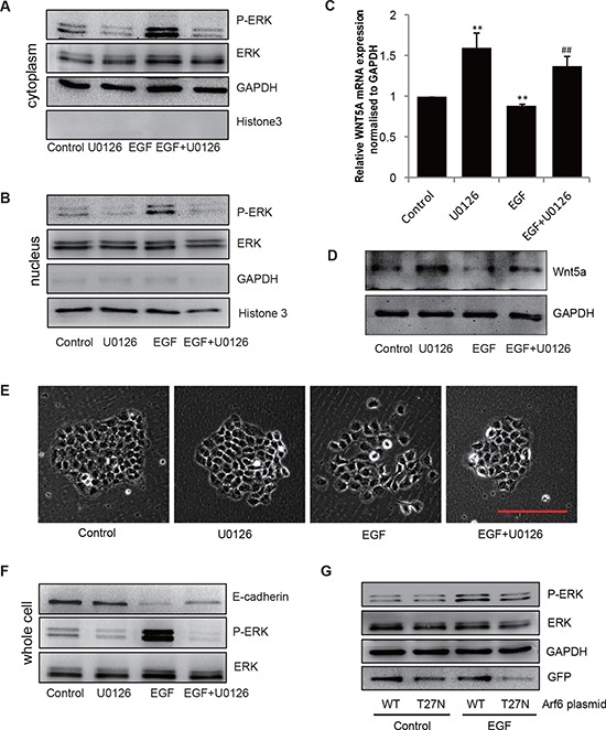
Figure 5. EGF/Arf6 reduces Wnt5a expression and promotes cell EMT through P-ERK.

(A–F) Cells were incubated for 2 h in the absence or presence of 10 μmol/L U0126 prior to EGF treatment (20 ng/mL for 48 h), the extracts of (A) cytoplasm and (B) nucleus were subjected to immunoblotting analysis to detect P-ERK. GAPDH or Histone 3 was as control for cytoplasm or nucleus part. (C) Total mRNA or (D) protein extracts for Wnt5a were analyzed by qPCR and immunoblotting. GAPDH was used as control. **P < 0.01 in the cultures with EGF or U0126 relative to the cultures without EGF. ##P < 0.01 in the cultures with EGF plus U0126 relative to the cultures with EGF alone. (E) The cell images were captured by phase-contrast microscopy. Scale bar, 100 μm. (F) The extracts of whole cell protein were subjected to immunoblotting analysis to detect E-cadherin. (G) SGC-7901 cells transfected with either an empty vector or an Arf6-T27N expression vector were stimulated with 20 ng/mL EGF for 48 h and ERK activity was examined.
