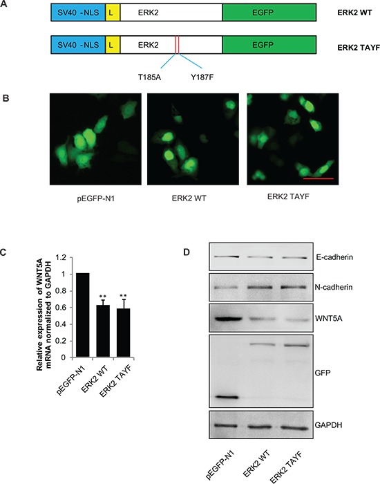
Figure 7. Wnt5a transcription and expression requires ERK2 nulear translocation, but not its phosphorylation.

(A) Schematic of the structure of the ERK2 and ERK2-TAYF mutant used for transfection. NLS: nulear localization signal. L:linker sequence. (B) Images of the cells transfected with pEGFP-N1, pEGFP-ERK2, and the pEGFP-ERK2-TAYF mutant. Scale bar, 50 μm. (C) Total mRNA extracts for Wnt5a were analyzed by qPCR and (D) total protein extracts for Wnt5a, E-cadherin and N-cadherin were analyzed by immunoblotting. **P < 0.01 in the cells transfected with the ERK2 and ERK2-TAYF mutant relative to the cells transfected with empty vector.
