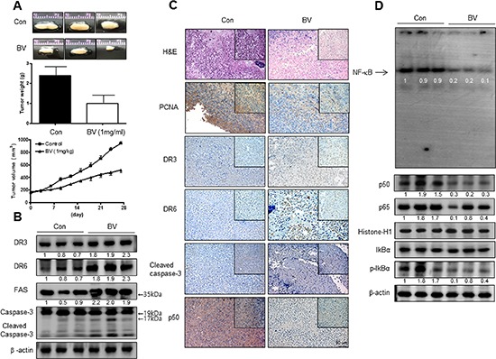
Figure 1. Anti-tumor activity of BV in cervical cancer xenograft.

Growth inhibition (as assessed by tumor volume) of subcutaneously transplanted CA Ski xenografts mice treated with BV (1 mg/kg/ two times a week) for 4weeks. Xenografted mice (n = 10) were administrated intraperitoneally with saline (1 ml/kg) or BV (1 mg/kg). Tumor burden was measured once per week using a caliper, and calculated volume length (mm) × width (mm) × height (mm)/2. Tumor weight and volume are presented as means ± S.D. (A). The expression of apoptotic proteins was detected by western blotting using specific antibodies; DR3, DR6, FAS, cleaved caspase-3 (B). β-actin protein was used an internal control. Immunohistochemistry was used to determine expression levels of H&E, PCNA, DR3, DR6, p50 in nude mice xenograft tissues by the different treatments as described in the Materials and Methods Section (C). NF-κB activity in tumor tissues (D). All values represent mean ± SD from five animal tumor sections. *P < 0.05 indicates significantly different from the control group.
